Anatomy Diagram Rib Area : Human Stomach Anatomy Diagram | Human Anatomy Body Picture ... - If you have mastered the anatomy of the arm, you know that the big, bulging biceps is positioned anteriorly (top of the image).
Anatomy Diagram Rib Area : Human Stomach Anatomy Diagram | Human Anatomy Body Picture ... - If you have mastered the anatomy of the arm, you know that the big, bulging biceps is positioned anteriorly (top of the image).. Go back to the diaphragm area and use a scalpel to cut the wall of the body cavity away from the diaphragm. Gross anatomy of the human female reproductive system 12. The salivary glands (element 30 in numbered diagram) in an insect's mouth produce saliva. At t11 and t12, the ribs do not attach and are so are called floating ribs. the thoracic spine's range of motion is limited due to the many rib/vertebrae connections and the long spinous processes. Identify the female reproductive system structures described below:
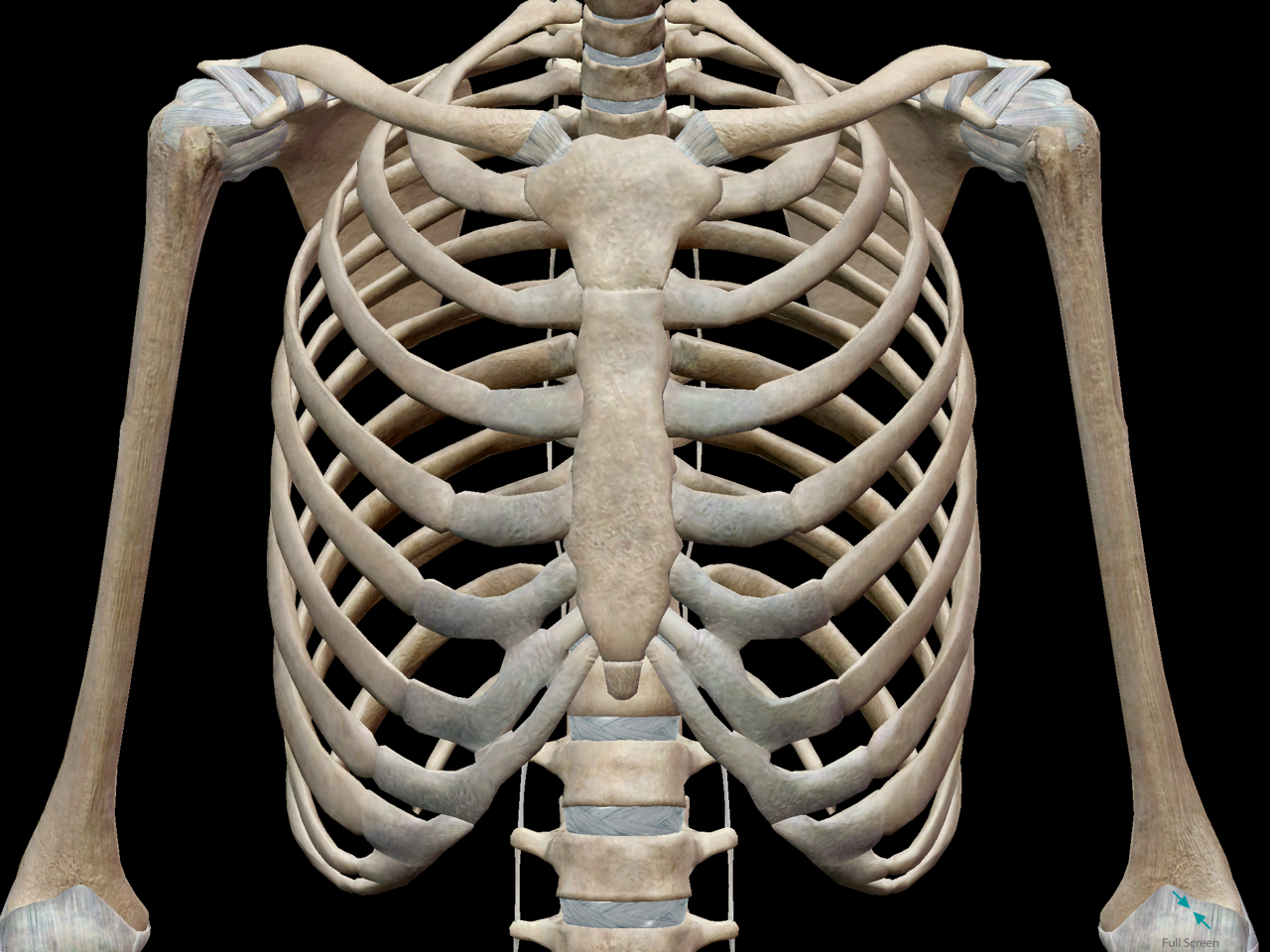
Oct 29, 2020 · the second intercostal space can be palpated on either side of this projection and is the location for auscultation of the pulmonary and aortic area on left and right respectively. "fertilized egg" typically formed here Learn vocabulary, terms, and more with flashcards, games, and other study tools. Identify the female reproductive system structures described below: The rib cage is the arrangement of ribs attached to the vertebral column and sternum in the thorax of most vertebrates that encloses and protects the vital organs such as the heart, lungs and great vessels.
Thorax Anterior view of human body - Biology Forums ... from i.pinimg.com The diaphragm should remain intact, but now the rib cage can be pulled back and pinned to the pan, exposing the thoracic cavity. The salivary ducts lead from the glands to the reservoirs and then forward through the head to an opening called the salivarium, located behind the hypopharynx. By moving its mouthparts (element 32 in numbered diagram) the insect can mix its food with saliva. If you have mastered the anatomy of the arm, you know that the big, bulging biceps is positioned anteriorly (top of the image). Learn vocabulary, terms, and more with flashcards, games, and other study tools. May 31, 2021 · cross section through the biceps brachii muscle: Gross anatomy of the human female reproductive system 12. On the diagram below of a frontal section of a portion of the female reproductive system, identify all indicated structures.
On the diagram below of a frontal section of a portion of the female reproductive system, identify all indicated structures.
Sep 10, 2019 · the rib cage is joined to the thoracic vertebrae. The diaphragm should remain intact, but now the rib cage can be pulled back and pinned to the pan, exposing the thoracic cavity. May 31, 2021 · cross section through the biceps brachii muscle: The rib cage is the arrangement of ribs attached to the vertebral column and sternum in the thorax of most vertebrates that encloses and protects the vital organs such as the heart, lungs and great vessels. In this position, a person is standing upright with the lower limbs together or slightly apart, feet flat on the floor and facing forward, upper limbs at the sides with the palms facing forward and thumbs pointing away from the body, and head and eyes directed. At t11 and t12, the ribs do not attach and are so are called floating ribs. the thoracic spine's range of motion is limited due to the many rib/vertebrae connections and the long spinous processes. Identify the female reproductive system structures described below: "fertilized egg" typically formed here If you have mastered the anatomy of the arm, you know that the big, bulging biceps is positioned anteriorly (top of the image). Learn vocabulary, terms, and more with flashcards, games, and other study tools. Gross anatomy of the human female reproductive system 12. The standard anatomical position is agreed upon by the international medical community. On the diagram below of a frontal section of a portion of the female reproductive system, identify all indicated structures.
The salivary ducts lead from the glands to the reservoirs and then forward through the head to an opening called the salivarium, located behind the hypopharynx. On the diagram below of a frontal section of a portion of the female reproductive system, identify all indicated structures. Notice how manubrium articulates with the clavicle via the clavicular notches and with the first rib at its lateral surface. The standard anatomical position is agreed upon by the international medical community. At t11 and t12, the ribs do not attach and are so are called floating ribs. the thoracic spine's range of motion is limited due to the many rib/vertebrae connections and the long spinous processes.
3D Skeletal System: 7 Interesting Facts about the Thoracic ... from cdn2.hubspot.net Learn vocabulary, terms, and more with flashcards, games, and other study tools. The diaphragm should remain intact, but now the rib cage can be pulled back and pinned to the pan, exposing the thoracic cavity. The standard anatomical position is agreed upon by the international medical community. Segmental anatomy in adults, each lung is 25cm to 30cm long, with the right lung being a little larger than the left lung as the former has three lobes while the latter only has two. May 31, 2021 · cross section through the biceps brachii muscle: "fertilized egg" typically formed here Make the last two incisions to expose the neck area. Sep 02, 2017 · it is the wide depressed area located just a little higher than the center of the medial surface of the lungs 11.
Gross anatomy of the human female reproductive system 12.
May 31, 2021 · cross section through the biceps brachii muscle: Notice how manubrium articulates with the clavicle via the clavicular notches and with the first rib at its lateral surface. Make the last two incisions to expose the neck area. Go back to the diaphragm area and use a scalpel to cut the wall of the body cavity away from the diaphragm. Oct 29, 2020 · the second intercostal space can be palpated on either side of this projection and is the location for auscultation of the pulmonary and aortic area on left and right respectively. Sep 10, 2019 · the rib cage is joined to the thoracic vertebrae. Learn vocabulary, terms, and more with flashcards, games, and other study tools. Gross anatomy of the human female reproductive system 12. Identify the female reproductive system structures described below: If you have mastered the anatomy of the arm, you know that the big, bulging biceps is positioned anteriorly (top of the image). Sep 02, 2017 · it is the wide depressed area located just a little higher than the center of the medial surface of the lungs 11. The salivary glands (element 30 in numbered diagram) in an insect's mouth produce saliva. In this position, a person is standing upright with the lower limbs together or slightly apart, feet flat on the floor and facing forward, upper limbs at the sides with the palms facing forward and thumbs pointing away from the body, and head and eyes directed.
Identify the female reproductive system structures described below: The salivary ducts lead from the glands to the reservoirs and then forward through the head to an opening called the salivarium, located behind the hypopharynx. The standard anatomical position is agreed upon by the international medical community. The salivary glands (element 30 in numbered diagram) in an insect's mouth produce saliva. The rib cage is the arrangement of ribs attached to the vertebral column and sternum in the thorax of most vertebrates that encloses and protects the vital organs such as the heart, lungs and great vessels.
Illustration Human Chest Ribs And Organs #8251677 Framed ... from www.mediastorehouse.com Make the last two incisions to expose the neck area. Notice how manubrium articulates with the clavicle via the clavicular notches and with the first rib at its lateral surface. The standard anatomical position is agreed upon by the international medical community. Sep 02, 2017 · it is the wide depressed area located just a little higher than the center of the medial surface of the lungs 11. Segmental anatomy in adults, each lung is 25cm to 30cm long, with the right lung being a little larger than the left lung as the former has three lobes while the latter only has two. In this position, a person is standing upright with the lower limbs together or slightly apart, feet flat on the floor and facing forward, upper limbs at the sides with the palms facing forward and thumbs pointing away from the body, and head and eyes directed. Standard anatomical position in humans. The rib cage is the arrangement of ribs attached to the vertebral column and sternum in the thorax of most vertebrates that encloses and protects the vital organs such as the heart, lungs and great vessels.
On the diagram below of a frontal section of a portion of the female reproductive system, identify all indicated structures.
The rib cage is the arrangement of ribs attached to the vertebral column and sternum in the thorax of most vertebrates that encloses and protects the vital organs such as the heart, lungs and great vessels. Identify the female reproductive system structures described below: Gross anatomy of the human female reproductive system 12. Notice how manubrium articulates with the clavicle via the clavicular notches and with the first rib at its lateral surface. Standard anatomical position in humans. Make the last two incisions to expose the neck area. The salivary ducts lead from the glands to the reservoirs and then forward through the head to an opening called the salivarium, located behind the hypopharynx. May 31, 2021 · cross section through the biceps brachii muscle: The diaphragm should remain intact, but now the rib cage can be pulled back and pinned to the pan, exposing the thoracic cavity. The salivary glands (element 30 in numbered diagram) in an insect's mouth produce saliva. The standard anatomical position is agreed upon by the international medical community. Sep 10, 2019 · the rib cage is joined to the thoracic vertebrae. In this position, a person is standing upright with the lower limbs together or slightly apart, feet flat on the floor and facing forward, upper limbs at the sides with the palms facing forward and thumbs pointing away from the body, and head and eyes directed.


0 Komentar